
Effects of Malaysian Kelulut Honey on Osteoblastic Proliferation of MC3T3-E1 Cells
DOI:
https://doi.org/10.54987/jobimb.v13i1.1084Keywords:
Malaysian Kelulut Honey, Osteoblasts, MC3T3-E1 Cells, Proliferation, Cell viabilityAbstract
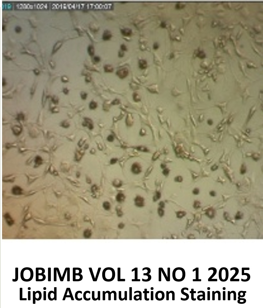
Malaysian Kelulut Honey (MKH) is a well-recognized Malaysian honey and has gained widespread popularity due to its ability to promote health. Numerous scientific studies have been conducted to prove the health benefits of MKH; however, its bone-protective effects have not yet been well-studied. Osteoblasts are vital cells for bone formation; thus, it is important to study the effect of MKH on osteoblasts for a better understanding of bone formation. This study was conducted to evaluate the effects of MKH on the proliferation of bone-forming cells using MC3T3-E1 cells. The cytotoxicity and proliferation of MKH on MC3T3-E1 cells were assessed by the 3-(4,5-dimethylthiazol-2-yl)-2,5-diphenyltetrazolium (MTT) assay and the bromodeoxyuridine (BrdU) assay. The cells were treated with MKH at various concentrations ranging from 0.001% v/v to 10.0% v/v for 24, 48, and 72 hours. The results demonstrated that MKH at 0.01% v/v significantly enhanced proliferation by 131.65% (p<0.001) after 24 hours compared to the negative control. However, cell viability and proliferation decreased in a dose-dependent manner at concentrations ≥0.05% v/v MKH, with 10.0% v/v MKH showing notable reduction in proliferation by over 40% (p<0.001) across all time points. Besides that, MKH concentrations below 0.1% v/v were non-cytotoxic and promoted osteoblast growth, indicating a limited effective range. These results suggest that MKH at low concentrations enhances osteoblast proliferation, supporting its potential as a natural agent for bone health.
References
Thu HE, Mohamed IN, Hussain Z, Shuid AN. Exploring molecular mechanism of bone-forming capacity of Eurycoma longifolia: Evidence of enhanced expression of bone-related biomarkers. J Ayurveda Integr Med. 2018;9(4):272-80.
Florencio-Silva R, da Silva Sasso GR, Sasso-Cerri E, Jesus Simões M, Sérgio Cerri P. Biology of Bone Tissue: Structure, Function, and Factors That Influence Bone Cells. Immuno-Anal Biol Spéc. 2015;2015(6):17.
Kenkre JS, Bassett JHD. The bone remodelling cycle. Ann Clin Biochem. 2018;55(3):308-27.
Crous A, Abrahamse H. The Signalling Effects of Photobiomodulation on Osteoblast Proliferation, Maturation and Differentiation: A Review. Stem Cell Rev Rep. 2021;1570-89.
Ralston SH. Bone structure and metabolism. Med U K. 2017;45(9):560-4.
Tu KN, Lie JD, Wan CKV, Cameron M, Austel AG, Nguyen JK, et al. Osteoporosis: A review of treatment options. P T. 2018;43(2):92-104.
Skjødt MK, Frost M, Abrahamsen B. Side effects of drugs for osteoporosis and metastatic bone disease. Br J Clin Pharmacol. 2019;85(6):1063-71.
Oryan A, Alemzadeh E, Moshiri A. Biological properties and therapeutic activities of honey in wound healing: A narrative review and meta-analysis. J Tissue Viability. 2016;25(2):98-118.
Rao PV, Krishnan KT, Salleh N, Gan SH. Biological and therapeutic effects of honey produced by honey bees and stingless bees: A comparative review. Braz J Pharmacogn. 2016;26(5):657-64.
Guan M, Pan D, Zhang M, Leng X, Yao B. The Aqueous Extract of Eucommia Leaves Promotes Proliferation, Differentiation, and Mineralization of Osteoblast-Like MC3T3-E1 Cells. Evid Based Complement Alternat Med. 2021;2021.
Wang D, Christensen K, Chawla K, Xiao G, Krebsbach PH, Franceschi RT. Isolation and characterization of MC3T3-E1 preosteoblast subclones with distinct in vitro and in vivo differentiation/mineralization potential. J Bone Miner Res. 1999;14(6):893-903.
Devasvaran K, Tan JJ, Ng CT, Fong LY, Yong YK. Malaysian Tualang Honey Inhibits Hydrogen Peroxide-Induced Endothelial Hyperpermeability. Oxid Med Cell Longev. 2019;2019.
Khalid AFBA, Ng CT, Fong LY, Tan JJ, Yakop FB, Kamaruddin NAB, et al. Malaysian Tualang Honey Suppresses the Angiogenic Events in Endothelial Cells Induced by Vascular Endothelial Growth Factor. Sains Malays. 2022 Mar 1;51(3):815-22.
Mosmann TR. Rapid colorimetric assay for cellular growth and survival: application to proliferation and cytotoxicity assays. J Immunol Methods. 1983;65 1-2:55-63.
Gratzner H G. Monoclonal Antibody to 5-Bromo-and 5-Iododeoxyuridine: A New Reagent for Detection of DNA Replication [Internet]. 1982 p. 474-5. (Science). Available from: 10.1126/science.7123245
Nordin A, Chowdhury SR, Saim AB, Idrus RBH. Effect of kelulut honey on the cellular dynamics of tgf?-induced epithelial to mesenchymal transition in primary human keratinocytes. Int J Environ Res Public Health. 2020;17(9).
Hazirah H, Yasmin AM, Abdul Karim N. Antioxidant Properties of Stingless Bee Honey and Its Effect on the Viability of Lymphoblastoid Cell Line. Med Health. 2019;14(1):91-105.
Ooi TC, Yaacob M, Rajab NF, Shahar S, Sharif R. The stingless bee honey protects against hydrogen peroxide-induced oxidative damage and lipopolysaccharide-induced inflammation in vitro. Saudi J Biol Sci. 2021;28(5):2987-94.
Yu LX, Taib H, Berahim Z, Ahmad A, Zainuddin SLA. The effect of Tualang honey on human periodontal ligament fibroblast proliferation and alkaline phosphatase level. Sains Malays. 2015;44(7):1021-5.
Kannan TP, Ali AQ, Abdullah SF, Ahmad A. Evaluation of Tualang honey as a supplement to fetal bovine serum in cell culture. Food Chem Toxicol. 2009;47(7):1696-702.
Yusof AM, Abd Ghafar N, Kamarudin TA, Hui CK, Yusof YAM. Gelam honey potentiates ex vivo corneal keratocytes proliferation with desirable phenotype expression. BMC Complement Altern Med. 2016;16(1):1-9.
Zhang Z, Lai Q, Li Y, Xu C, Tang X, Ci J, et al. Acidic pH environment induces autophagy in osteoblasts. Sci Rep [Internet]. 2017 Apr 6 [cited 2025 July 16];7(1). Available from: https://www.nature.com/articles/srep46161
Abdelaziz AG, Nageh H, Abdalla MS, Abdo SM, Amer AA, Loutfy SA, et al. Development of polyvinyl alcohol nanofiber scaffolds loaded with flaxseed extract for bone regeneration: phytochemicals, cell proliferation, adhesion, and osteogenic gene expression. Front Chem. 2024;12.
Xing L, Li Y, Li W, Liu R, Geng Y, Ma W, et al. Expression of RUNX2/LAPTM5 in the Induction of MC3T3-e1 Mineralization and Its Possible Relationship with Autophagy. Tissue Eng Regen Med. 2022 Dec 1;19(6):1223-35.
Jaara HS, Torres S. Mitochondrial ROS, a trigger for mitochondrial dysfunction and inflammasome activation and a therapeutic target in liver diseases. Explor Dig Dis. 2024 Dec 10;3(6):474-503.
Downloads
Published
Issue
Section
License
Copyright (c) 2025 Nur Najihah Ahmad Puat, Yong Yoke Keong, Nizar Abd Manan

This work is licensed under a Creative Commons Attribution 4.0 International License.
Authors who publish with this journal agree to the following terms:
- Authors retain copyright and grant the journal right of first publication with the work simultaneously licensed under a Creative Commons Attribution License (http://creativecommons.org/licenses/by/4.0) that allows others to share the work with an acknowledgement of the work's authorship and initial publication in this journal.
- Authors are able to enter into separate, additional contractual arrangements for the non-exclusive distribution of the journal's published version of the work (e.g., post it to an institutional repository or publish it in a book), with an acknowledgement of its initial publication in this journal.
- Authors are permitted and encouraged to post their work online (e.g., in institutional repositories or on their website) prior to and during the submission process, as it can lead to productive exchanges, as well as earlier and greater citation of published work (See The Effect of Open Access).
