Characterization of the Growth of Pseudomonas sp. strain DrY135 on Acrylamide
DOI:
https://doi.org/10.54987/jemat.v10i2.734Keywords:
Acrylamide, Pseudomonas sp., Bioremediation, Biodegradation, CharacterizationAbstract
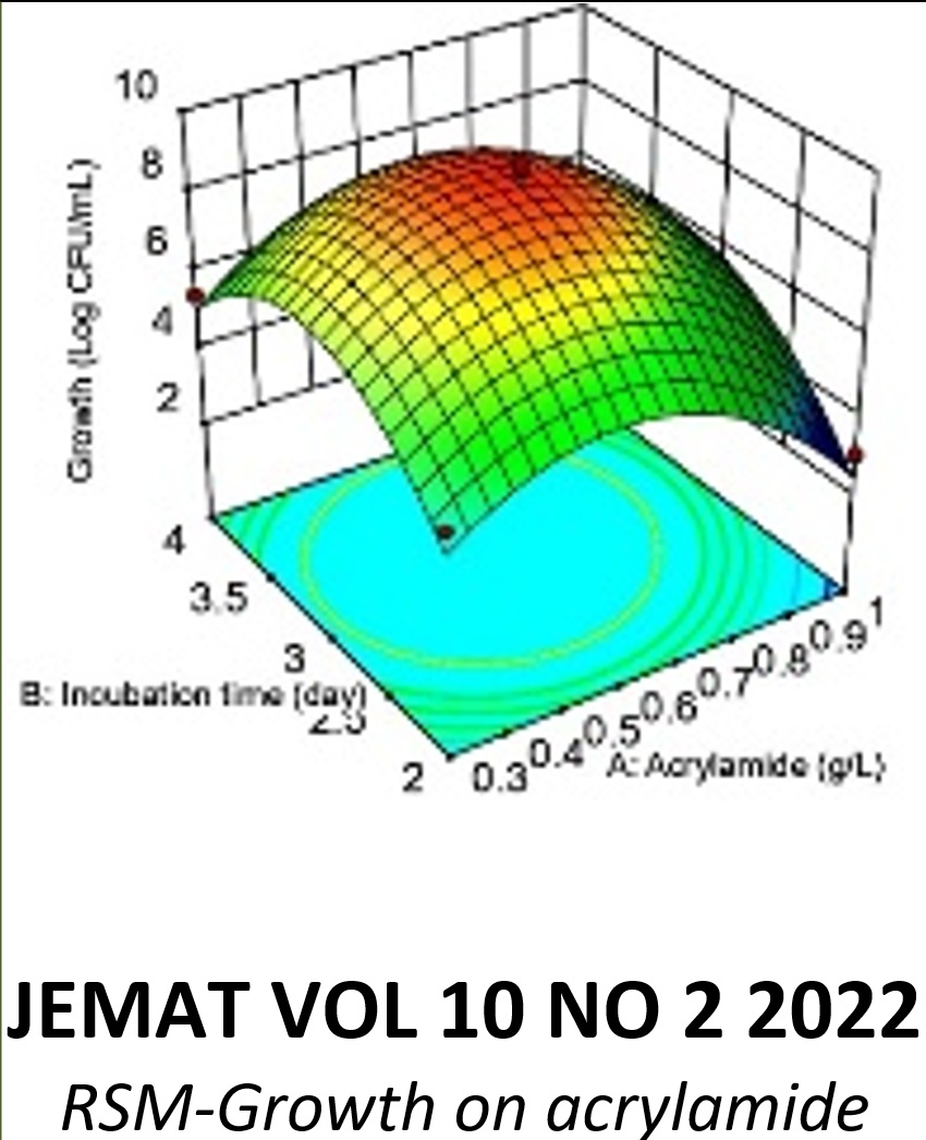
This study investigated the growth properties of a molybdenum-reducing bacteria previously isolated for its ability to break down amides. The bacterial growth range is 500–1000 mg/L, 6.5–8.0 pH, and 30–35 °C. The presence of hazardous heavy metals such as mercury, silver, and copper impeded this bacterium's development on acrylamide. The protracted lag phase seen when growing on acrylamide demonstrates the compound's severe growth inhibition. This bacterium has the potential to be an effective acrylamide bioremediation agent due to its greater tolerance for acrylamide than other acrylamide-degrading bacteria identified in the scientific literature. The influence of initial pH on bacterial growth at room temperature indicates that the optimal pH range lies between 6.5 and 8.0. The ideal temperature range for plant growth was between 30 and 35 oC. In a series of experiments utilizing a starting concentration of 1% (w/v) of various organic carbon sources, it was determined that glucose supported the most cellular growth on acrylamide, followed by sucrose, fructose, mannose, and citrate, in descending order of efficiency, whereas mannitol did not support growth. Doses of 300 and 500 mg/L of acrylamide stimulated the most rapid growth expansion, but concentrations of 1500 mg/L and above completely halted development. Copper (Cu), lead (Pb), cadmium (Cd), chromium (Cr), and mercury (Hg) were investigated at a concentration of 2 ppm. Mercury hindered growth by 71 percent, copper by 72 percent, and cadmium by 52 percent, according to our findings. There was a linear association between the acrylamide content and the delay before this bacterium began to develop. A lag time of one to three days was found when the acrylamide content grew from 100 to 1,500 mg/L. As quantities of acrylamide increased, so did the maximal growth rate, indicating an overall pattern of increasing toxicity.
References
Mottram, DS, Wedzicha BL, Dobson AT. Acrylamide is formed in the Maillard reaction. Nature. 2002;419:448-9.
Zamora R, Delgado RM, Hidalgo FJ. Strecker aldehydes and ?-keto acids, produced by carbonyl-amine reactions, contribute to the formation of acrylamide. Food Chem. 2011;128(2):465-70.
Shukor MY, Gusmanizar N, Azmi NA, Hamid M, Ramli J, Shamaan NA, et al. Isolation and characterization of an acrylamide-degrading Bacillus cereus. J Enviromental Biol. 2009;30(1):57-64.
Sega GA, Valdivia Alcota RP, Tancongco CP, Brimer PA. Acrylamide binding to the DNA and protamine of spermiogenic stages in the mouse and its relationship to genetic damage. Mutat Res Mutagen Relat Subj. 1989 Aug 1;216(4):221-30.
Tyl RW, Friedman MA. Effects of acrylamide on rodent reproductive performance. Reprod Toxicol. 2003 Jan 1;17(1):1-13.
Yang HJ, Lee SH, Jin Y, Choi JH, Han CH, Lee MH. Genotoxicity and toxicological effects of acrylamide on reproductive system in male rats. J Vet Sci. 2005 Jun;6(2):103-9.
Backer LC, Dearfield KL, Erexson GL, Campbell JA, Westbrook?Collins B, Allen JW. The effects of acrylamide on mouse germ-line and somatic cell chromosomes. Environ Mol Mutagen. 1989;13(3):218-26.
Spencer P, Schaumburg HH. Nervous system degeneration produced by acrylamide monomer. Environ Health Perspect. 1975 Jun 1;11:129-33.
Hagmar L, Törnqvist M, Nordander C, Rosén I, Bruze M, Kautiainen A, et al. Health effects of occupational exposure to acrylamide using hemoglobin adducts as biomarkers of internal dose. Scand J Work Environ Health. 2001;27(4):219-26.
Igisu H, Goto I, Kawamura Y, Kato M, Izumi K. Acrylamide encephaloneuropathy due to well water pollution. J Neurol Neurosurg Psychiatry. 1975;38(6):581-4.
Eikmann T, Herr C. How dangerous is actually acrylamide exposure for the population. Umweltmed Forsch Prax. 2002;7(6):307-8.
Pruser KN, Flynn NE. Acrylamide in health and disease. Front Biosci - Sch. 2011;3 S(1):41-51.
Pennisi M, Malaguarnera G, Puglisi V, Vinciguerra L, Vacante M, Malaguarnera M. Neurotoxicity of acrylamide in exposed workers. Int J Environ Res Public Health. 2013;10(9):3843-54.
Wampler DA, Ensign SA. Photoheterotrophic metabolism of acrylamide by a newly isolated strain of Rhodopseudomonas palustris. Appl Environ Microbiol. 2005;71(10):5850-7.
Buranasilp K, Charoenpanich J. Biodegradation of acrylamide by Enterobacter aerogenes isolated from wastewater in Thailand. J Environ Sci. 2011;23(3):396-403.
Charoenpanich J, Tani A. Proteome analysis of acrylamide-induced proteins in a novel acrylamide-degrader Enterobacter aerogenes by 2D electrophoresis and MALDI-TOF-MS. Chiang Mai Univ J Nat Sci. 2014;13(1):11-22.
Gusmanizar N, Shukor Y, Ramli J, Syed MA. Isolation and characterization of an acrylamide-degrading Burkholderia sp. strain DR.Y27. J Ris Kim. 2015 Feb 11;2(1):34.
Yu F, Fu R, Xie Y, Chen W. Isolation and characterization of polyacrylamide-degrading bacteria from dewatered sludge. Int J Environ Res Public Health. 2015;12(4):4214-30.
Bedade DK, Singhal RS. Biodegradation of acrylamide by a novel isolate, Cupriavidus oxalaticus ICTDB921: Identification and characterization of the acrylamidase produced. Bioresour Technol. 2018 Aug 1;261:122-32.
Aisami A, Gusmanizar N. Characterization of an acrylamide-degrading bacterium isolated from hydrocarbon sludge. Bioremediation Sci Technol Res. 2019 Dec 28;7(2):15-9.
Othman AR, Rahim MBHA. Modelling the Growth Inhibition Kinetics of Rhodotorula sp. strain MBH23 (KCTC 11960BP) on Acrylamide. Bioremediation Sci Technol Res. 2019 Dec 28;7(2):20-5.
Rusnam, Gusmanizar N. An Acrylamide-degrading Bacterial Consortium Isolated from Volcanic Soil. J Biochem Microbiol Biotechnol. 2021 Dec 31;9(2):19-24.
Rusnam, Gusmanizar N. Characterization of An Acrylamide-degrading Bacterium Isolated from Volcanic Soil. J Environ Bioremediation Toxicol. 2022 Aug 5;5(1):32-7.
Yakasai MH, Abd Rahman MF, Abd Rahim MBH, Khayat ME, Shamaan NA, Shukor MY. Isolation and characterization of a metal-reducing Pseudomonas sp. strain 135 with amide-degrading capability. Bioremediation Sci Technol Res. 2017;5(2):32-8.
Svensson K, Abramsson L, Becker W, Glynn A, Hellenäs KE, Lind Y, et al. Dietary intake of acrylamide in Sweden. Food Chem Toxicol. 2003;41(11):1581-6.
Smith EA, Prues SL, Oehme FW. Environmental degradation of polyacrylamides. 1. Effects of artificial environmental conditions: Temperature, light, and pH. Ecotoxicol Environ Saf. 1996;35(2):121-35.
Rogacheva SM, Ignatov OV. The respiratory activity of Rhodococcus rhodochrous M8 cells producing nitrile-hydrolyzing enzymes. Appl Biochem Microbiol. 2001;37(3):282-6.
Jonston JJ, Borden RC, Barlaz MA. Anaerobic biodegradation of alkylbenzenes and trichloroethylene in aquifer sediment down gradient of a sanitary landfill. J Contam Hydrol. 1996;23(4):263-83.
Baek SH, Kim KH, Yin CR, Jeon CO, Im WT, Kim KK, et al. Isolation and characterization of bacteria capable of degrading phenol and reducing nitrate under low-oxygen conditions. Curr Microbiol. 2003;47(6):462-6.
Egorova K, Trauthwein H, Verseck S. Purification and properties of an enantioselective and thermoactive amidase from the thermophilic actinomycete Pseudonocardia thermophila. Appl Microbiol Biotechnol. 2004;38-45.
Rahim MBH, Syed MA, Shukor MY. Isolation and characterization of an acrylamide-degrading yeast Rhodotorula sp . strain MBH23 KCTC 11960BP. J Basic Microbiol. 2012;52(5):573-81.
Rogacheva SM, Ignatov OV. The Respiratory Activity of Rhodococcus rhodochrous M8 Cells Producing Nitrile-Hydrolyzing Enzymes. Appl Biochem Microbiol. 2001;37(3):282-6.
Shukor MY, Ahmad SA, Nadzir MMM, Abdullah MP, Shamaan NA, Syed MA. Molybdate reduction by Pseudomonas sp . strain DRY2. J Appl Microbiol. 2010;108:2050-8.
Shen S min, Wan T jou, Hwang H yuan. Biocatalysis and Agricultural Biotechnology Enhancement of degradation of acrylamide coupled with salad oil by Pseudomonas aeruginosa DS-4 using incubation periods. Biocatal Agric Biotechnol. 2012;1(2):110-4.
Wakaizumi M, Yamamoto H, Fujimoto N, Ozeki K. Acrylamide degradation by filamentous fungi used in food and beverage industries. J Biosci Bioeng. 2009;108(5):391-3.
Cha M, Chambliss GH. Characterization of Acrylamidase Isolated from a Newly Isolated Acrylamide-Utilizing Bacterium , Ralstonia eutropha AUM-01. Curr Microbiol. 2011;671-8.
Shukor MY, Gusmanizar N, Ramli J, Shamaan NA, Maccormack WP, Syed MA. Isolation and characterization of an acrylamide-degrading Antarctic bacterium. J Enviromental Biol. 2009;30(1):107-12.
Amor L, Kennes C, Veiga MC. Kinetics of inhibition in the biodegradation of monoaromatic hydrocarbons in presence of heavy metals. Bioresour Technol. 2001 Jun 1;78(2):181-5.
Gopinath KP, Kathiravan MN, Srinivasan R, Sankaranarayanan S. Evaluation and elimination of inhibitory effects of salts and heavy metal ions on biodegradation of Congo red by Pseudomonas sp. mutant. Bioresour Technol. 2011;102(4):3687-93.
Roane TM, Josephson KL, Pepper IL. Dual-Bioaugmentation Strategy To Enhance Remediation of Cocontaminated Soil. Appl Environ Microbiol. 2001 Jul;67(7):3208-15.
Hettiarachchi GM, Pierzynski GM, Ransom MD. In situ stabilization of soil lead using phosphorus and manganese oxide. Environ Sci Technol. 2000;34(21):4614-9.
Babich H, Stotzky G. Effect of Cadmium on Fungi and on Interactions Between Fungi and Bacteria in Soil: Influence of Clay Minerals and pH. Appl Environ Microbiol. 1977 May;33(5):1059-66.
Kamel Z. Toxicity of cadmium to twoStreptomyces species as affected by clay minerals. Plant Soil. 1986 Jun 1;93(2):195-203.
Downloads
Published
Issue
Section
License
Copyright (c) 2022 Journal of Environmental Microbiology and Toxicology

This work is licensed under a Creative Commons Attribution 4.0 International License.
Authors who publish with this journal agree to the following terms:
- Authors retain copyright and grant the journal right of first publication with the work simultaneously licensed under a Creative Commons Attribution License (http://creativecommons.org/licenses/by/4.0) that allows others to share the work with an acknowledgement of the work's authorship and initial publication in this journal.
- Authors are able to enter into separate, additional contractual arrangements for the non-exclusive distribution of the journal's published version of the work (e.g., post it to an institutional repository or publish it in a book), with an acknowledgement of its initial publication in this journal.
- Authors are permitted and encouraged to post their work online (e.g., in institutional repositories or on their website) prior to and during the submission process, as it can lead to productive exchanges, as well as earlier and greater citation of published work (See The Effect of Open Access).
